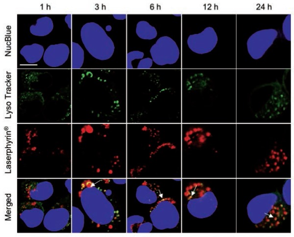
Figure 5:

Subcellular localization of Laserphyrin®. PC-3 cells were incubated in 3.75 mM Laserphyrin® solution for various incubation periods. Fluorescence images of nuclei (blue), lysosomes (green), and Laserphyrin® (red) were visualized by confocal microscopy. Distributions of Laserphyrin® coincided closely with those of lysosomes, indicating colocalization of Laserphyrin® with lysosomes. Scale bar, 50 µm.
