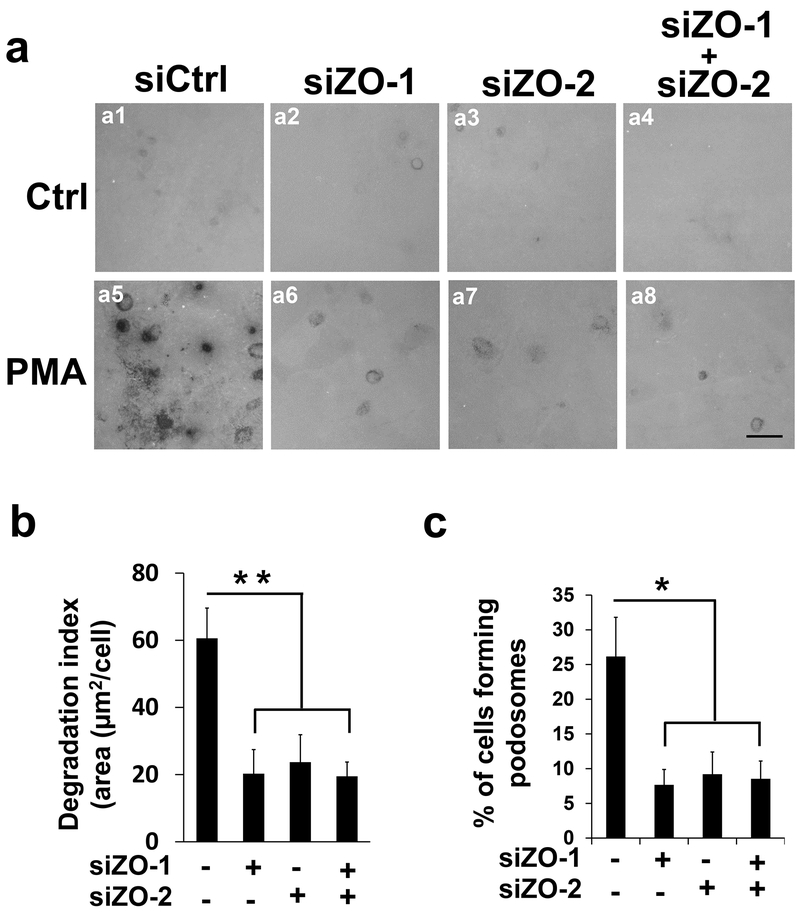
Figure 2. ZO-1 and ZO-2 are essential for podosome function in a non-redundant manner.
(a) SiRNAs against ZO-1 (siZO-1, 2 nM) or ZO-2 (siZO-2, 5 nM) were introduced in HBMECs singly (a2-3, and a6-7) or in combination (a4 and a8), followed by in situ extracellular matrix degradation assay in the absence (the first row) and presence of PMA treatment (the second row, 100 nM for 1 hour). Dark spots are caused by the loss of fluorescent signals from alexa488-labeled gelatin that was pre-coated onto the chamber slides. (b-c) At least 9 different fields (10X objectives) were selected to calculate the degradation index (b) and percentage of podosome formation (c) as described in Materials and Methods. n=3; scale bar: 10 µm. Data were represented as mean±s.e.m. *p<0.05, **p<0.01.

