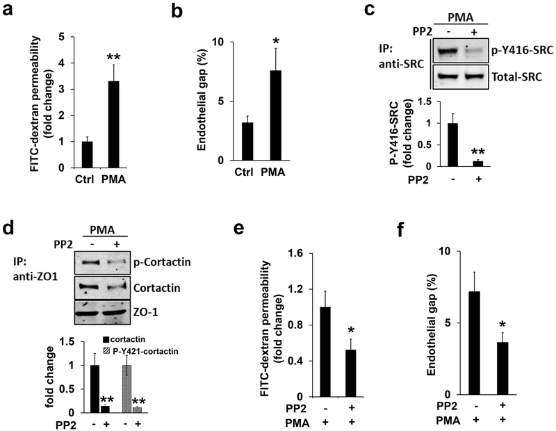
Figure 6. SRC kinase inhibition ameliorates PMA-induced endothelial barrier defect.
(a-b) FITC-dextran permeability assay (a) or endothelial gap formation assay (b) were applied to HMBECs in the absence (DMSO) and presence of PMA treatment, as described in Materials and Methods. Note that PMA treatment significantly induced endothelial barrier leakiness. (c-d) HBMECs pre-incubated with PP2 (10 µM, 1 hour) or DMSO (Ctrl) were further stimulated by PMA treatment (100 nM, 1 hour). Western blotting revealed that the level of p-Y416-SRC (c) or cortactin and p-Y421-cortactin co-immunoprecipitated by ZO-1 (d) was reduced, respectively. Total protein level of SRC or immunoprecipitated ZO-1 as loading control as indicated, and quantifications were shown in the bar graphs on the bottom (e-f). Inhibition of SRC kinase by PP2 treatment further attenuated PMA-induced endothelial barrier defects judged by the FITC-dextran permeability assay (e) and endothelial gap formation assay (f), respectively.

