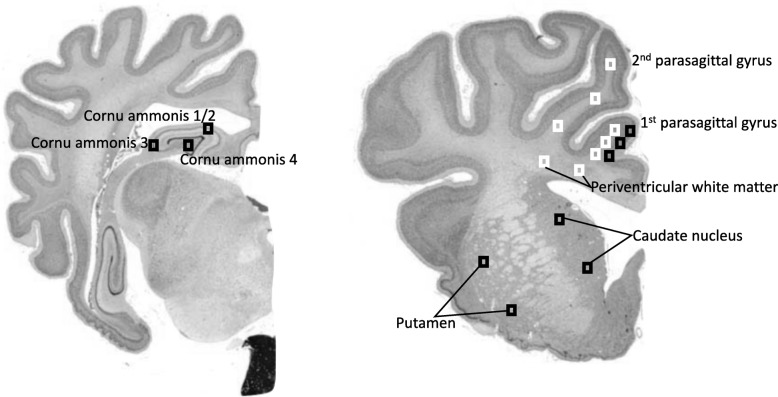
Fig. 2.
Schematic indicating fields sampled (regions of interest) for histological assessment. Left shows the cornu ammonis of the dorsal horn of the anterior hippocampus (CA 1–2, 3, 4) that was examined on sections taken 17 mm anterior to stereotaxic zero. Right shows brain regions of the forebrain used for analysis; these included the premotor cortex, caudate nucleus and putamen at the level of the mid-striatum, and periventricular and intragyral white matter from sections taken 23 mm anterior to stereotaxic zero. Black squares were sampled for assessment of neuronal survival within the first parasagittal gyri, caudate nucleus, and putamen. White squares were sampled for assessment of intragyral white matter within the first and second parasagittal gyri and the periventricular white matter. Image source: [31]

