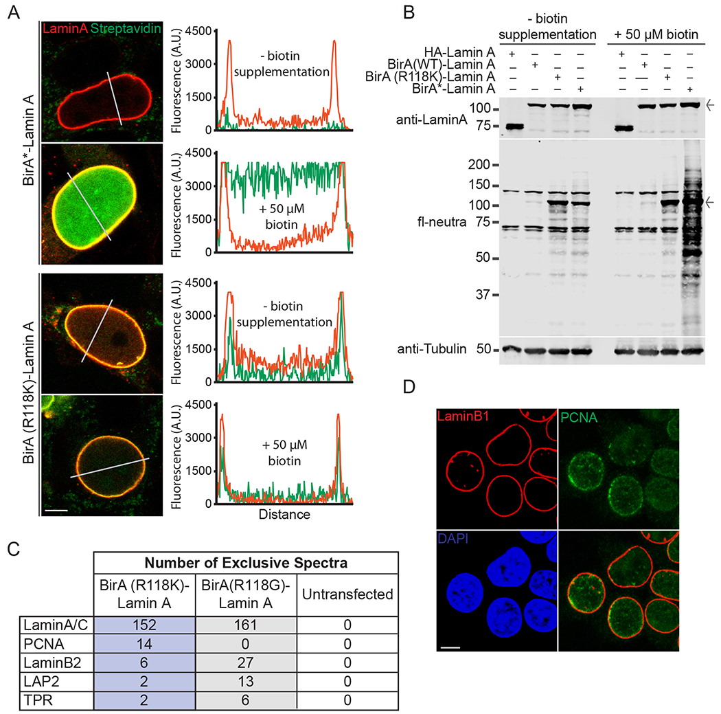
Fig. 6.

Cell-based experiments with BirA-Lamin A fusions. (A) Confocal microscopy showing the localization (red) and biotinylation (green) of BirA*-Lamin A and BirA(R118K)-Lamin A. HEK293T cells grown in the absence and presence of exogenous biotin (50 μM). Fluorescence intensities were measured across nuclei (white lines) and quantified (middle panels) (Scale bar 5 μm) (B) Whole cell lysate from HEK293T cells transfected with HA-Lamin A or BirA-Lamin A fusions with and without biotin supplementation (50 μM). Immunoblot of fl-neutra detection (arrow denotes BirA-Lamin A band) (C) Mass spectrometry results obtained from streptavidin pulldown of HEK293T lysate from cells expressing Lamin A-BirA(R118K) and Lamin A-BirA* in the absence of additional biotin supplementation. Shown are selected peptides derived from proteins known to be proximal to Lamin A in cells. A complete list of peptides from this analysis is provided (Table S3). (D) Localization of endogenous PCNA to the nuclear lamina by confocal fluorescence microscopy (Scale bar 5 μm). (For interpretation of the references to colour in this figure legend, the reader is referred to the web version of this article.)
