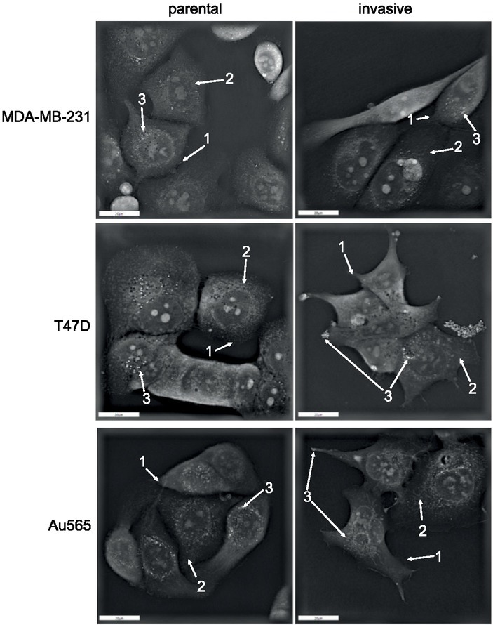
Figure 1.

Holographic microscopy of breast cancer cells. Parental and invasive MDA-MB-231, T47D, and Au565 cells were analysed for their morphology using 3D tomographic microscope as described in Materials and Methods. Arrow 1: filopodia; Arrow 2: mitochondria; Arrow 3: lipid droplets.
