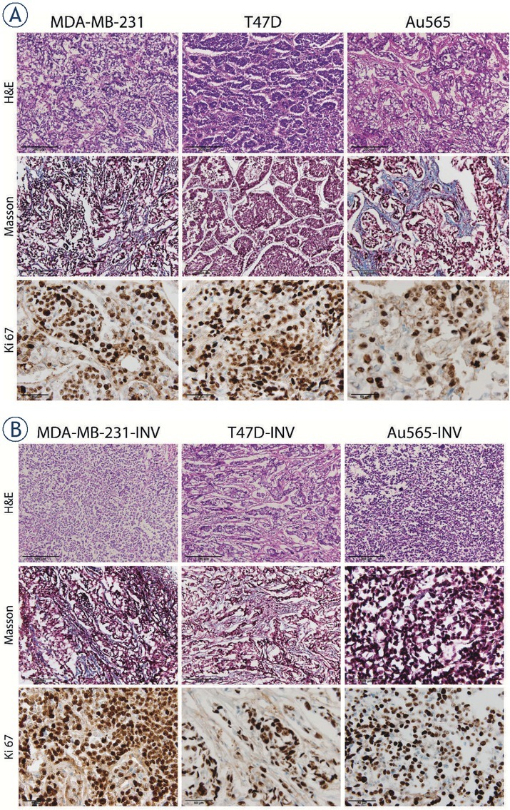
Figure 9.

Representative histological images of breast cancer xenografts. Tumors originating from parental (A) and invasive (B) MDA-MB-231, T47D and Au565 breast carcinoma cells were stained with H&E for evaluation of tumor morphology (200× magnification). Collagen (blue) was stained using Masson`s staining (200× magnification). Ki-67 proliferative tumor cells are stained brown (600× magnification).
