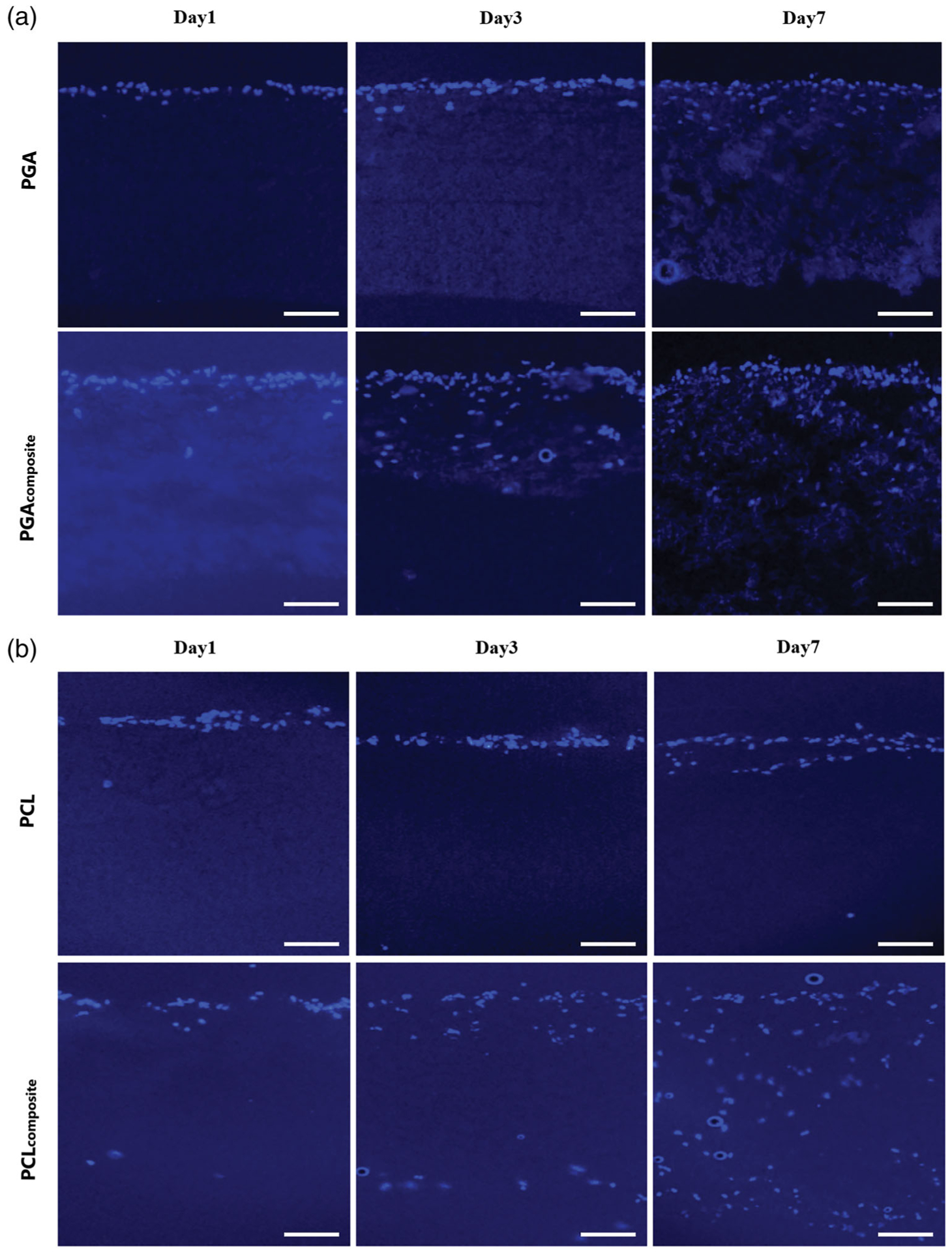
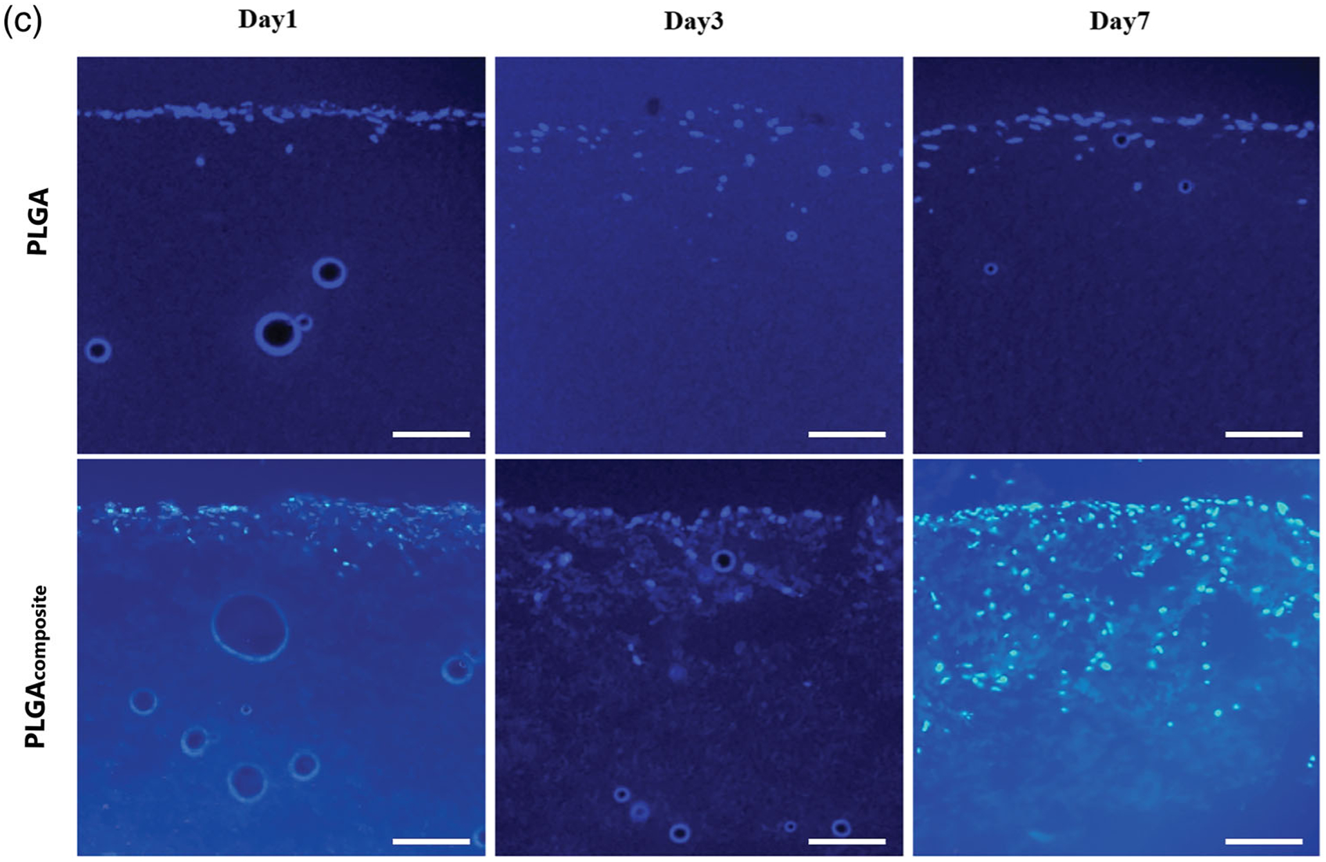
FIGURE 4.


The cross-section histology of the fibroblast cell infiltration into the electrospun and composite scaffolds with DAPI staining (cell nuclei) on a fluorescent microscope at day1, 3, and 7. (a) PGA, (B) PCL, (C) PLGA. Scale bar: 100 μm (all images)
