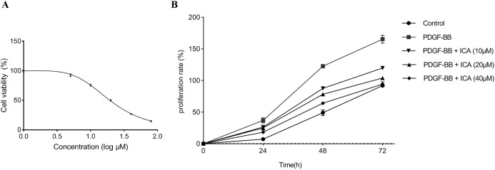
Figure 1. Cell viability was assessed in RPE cells by using the MTS assay.
(A) ICA treatments exerted an inhibitory effect on RPE cells without stimulation of PDGF-BB. After a series concentration of ICA (0, 1, 5, 10, 20, 40 and 80 μM) treatments for 48 h, the IC50 value of ICA was calculated using GraphPad Prism 8.0. (B) The cellular proliferating ability was assessed by the proliferation curve at 0, 24, 48 and 72 h after ICA treatment. Control: blank control group without PDGF-BB; PDGF-BB: 20 ng/ml PDGF-BB; PDGF-BB + ICA (10 µM): 20 ng/ml PDGF-BB + 10 µM ICA; PDGF-BB + ICA (20 µM): 20 ng/ml PDGF-BB + 20 µM ICA; PDGF-BB + ICA (40 µM): 20 ng/ml PDGF-BB + 40 µM ICA. Data are expressed as mean ± SD.

