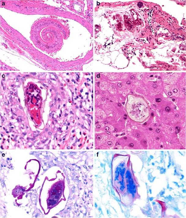
Fig. 10.

Intestinal schistosomiasis. Schistosoma mansoni worm residing in a mesenteric vein (a, ×100). Inert calcified elongated S. mansoni ova in rectal submucosa (b, ×200). Comparison of viable elongated S. mansoni ovum showing surrounding eosinophil-rich granulomatous inflammation (c, ×400) and smaller, ovoid S. mekongi ovum (in the liver) with small knob-like spine (d, ×400). S. mansoni ova showing PAS-positivity (e, ×400) and acid-fast staining of shell and spine (f, ×400)
