Fig. 5.

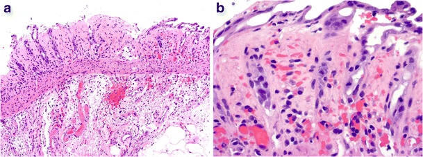
Enterohaemorrhagic Escherichia coli infection with mucosal erosion, prominent submucosal edema and fibrin thrombi (a, ×100). Note the withering crypts, lamina propria red cell extravasation and hyalinisation (b, ×400)

Enterohaemorrhagic Escherichia coli infection with mucosal erosion, prominent submucosal edema and fibrin thrombi (a, ×100). Note the withering crypts, lamina propria red cell extravasation and hyalinisation (b, ×400)