Fig. 1.

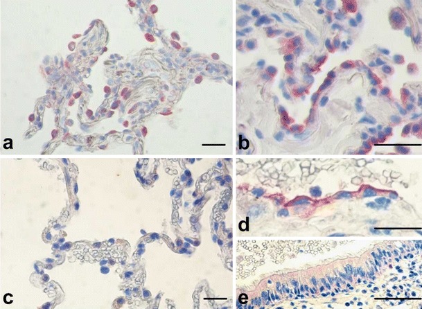
Immunohistochemical (IHC) staining of TLR2 in human lung tissue before (a) and after (b) in vitro infection with nontypeable H. influenzae. IHC of TLR9 in human lung tissue before (c) and after (d, AEC; and e bronchial epithelial cells) in vitro infection with nontypeable H. influenzae (scale bar 50μm)
