Fig. 3.

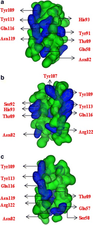
Monomers according to the assembly protocol: Seq1 (a), Seq2 (b) and Sim (c). Hydrophilic residues are highlighted in blue, hydrophobic residues in green. All models are drawn in a ‘Gaussian Contact’ illustration (MOE)

Monomers according to the assembly protocol: Seq1 (a), Seq2 (b) and Sim (c). Hydrophilic residues are highlighted in blue, hydrophobic residues in green. All models are drawn in a ‘Gaussian Contact’ illustration (MOE)