Figure 5.
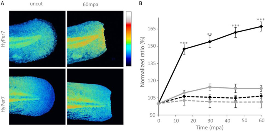
Monitoring H2O2 production with HyPer7 in zebrafish larvae after wounding. Zebrafish embryos were injected with HyPer7 or HyPer7-C121S mRNAs at the one-cell stage and tail fin amputation assay was performed on 48 hpf larvae. Images were taken before amputation and time lapse imaging was performed up to 60 min post-amputation (mpa). HyPer7 and HyPer7-C121S ratios were quantified at the amputation plane and normalized to the mean fluorescence of the uncut tail for each larvae. (A) Representative images of HyPer7 and HyPer7-C121S expressing larvae before and after amputation. Scale bar is 100µm. (B) Time course of HyPer7 (black lines) or HyPer7-C121S (gray lines) in the wounding site of the larvae tail fin (values ± SEM, N=2) experiments, n≥4 embryos/time point; **, P<0,01; ***, P<0,001). Uncut tails - dashed line.

