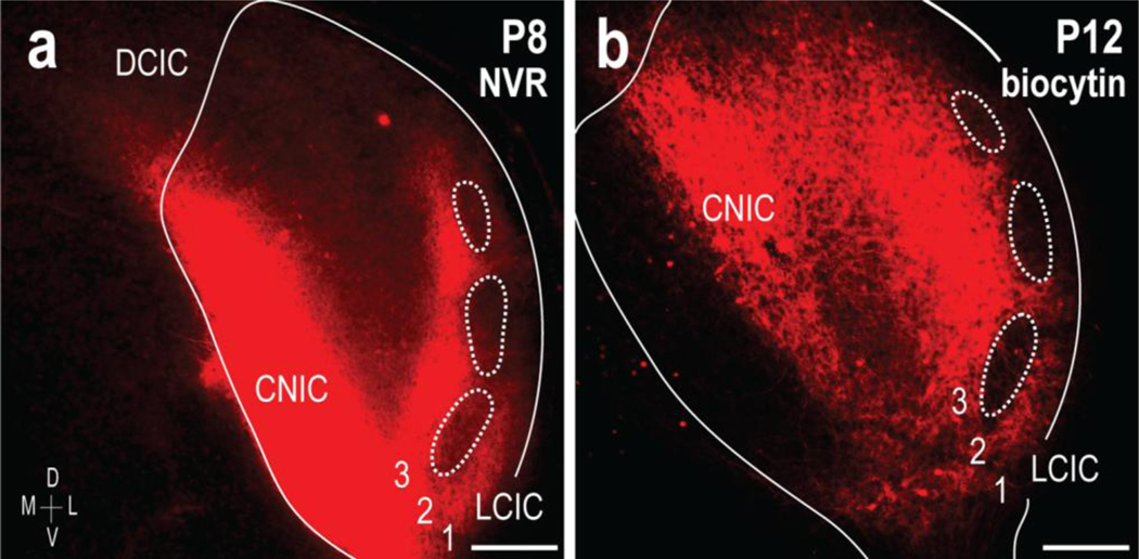
Fig 1.

Anterograde tracing approaches in early postnatal mouse IC. Placement of NeuroVue Red dye-soaked filter paper in the CNIC labels projections to the LCIC (a, right IC, ipsilateral input shown). Inputs preferentially target presumptive extramodular zones by P8, terminating throughout layers 1 and 3, as well as intermodular areas of layer 2. Similar CNIC placements of biocytin in living slice preparations yield comparable results, with extramodular projection patterns that avoid LCIC layer 2 patches (b, dashed contours). Unlike pilot lipophilic dye tracings in fixed tissue, this approach facilitates better visualization at later developmental stages after myelination (P12 shown in b), as well as being compatible with subsequent immunostaining protocols (see Figs. 8–10). Scale bars = 200 μm
