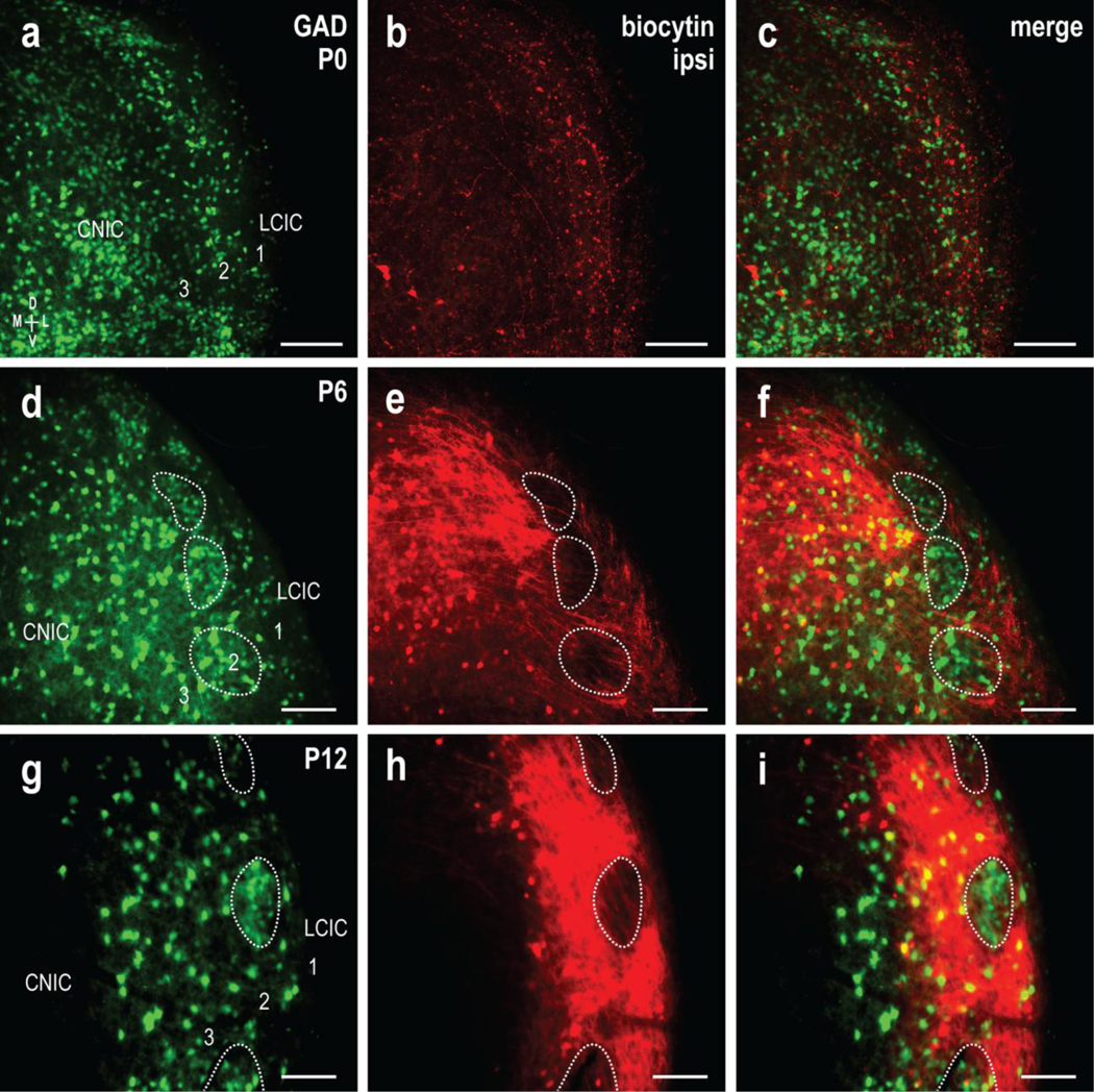
Fig 3.

Developmental segregation of the uncrossed CNIC projection from GAD-positive LCIC modular fields. GAD67-GFP (green) and biocytin labeling (red) of the ipsilateral input at P0 (a-c), P6 (d-f), and P12 (g-i). At birth, some CNIC fibers occupy aspects of the LCIC, although labeling was sparse and unorganized. With emerging compartments at P6, uncrossed inputs show a preference for extramodular zones, with few fibers evident in GAD-defined modules (d-f, dashed contours). By P12, labeled projection distributions were consistently robust and discretely mapped to extramodular zones, with prominent voids throughout layer 2 that coincided with GAD-positive modules (g-i, dashed contours). Scale bars = 100μm
