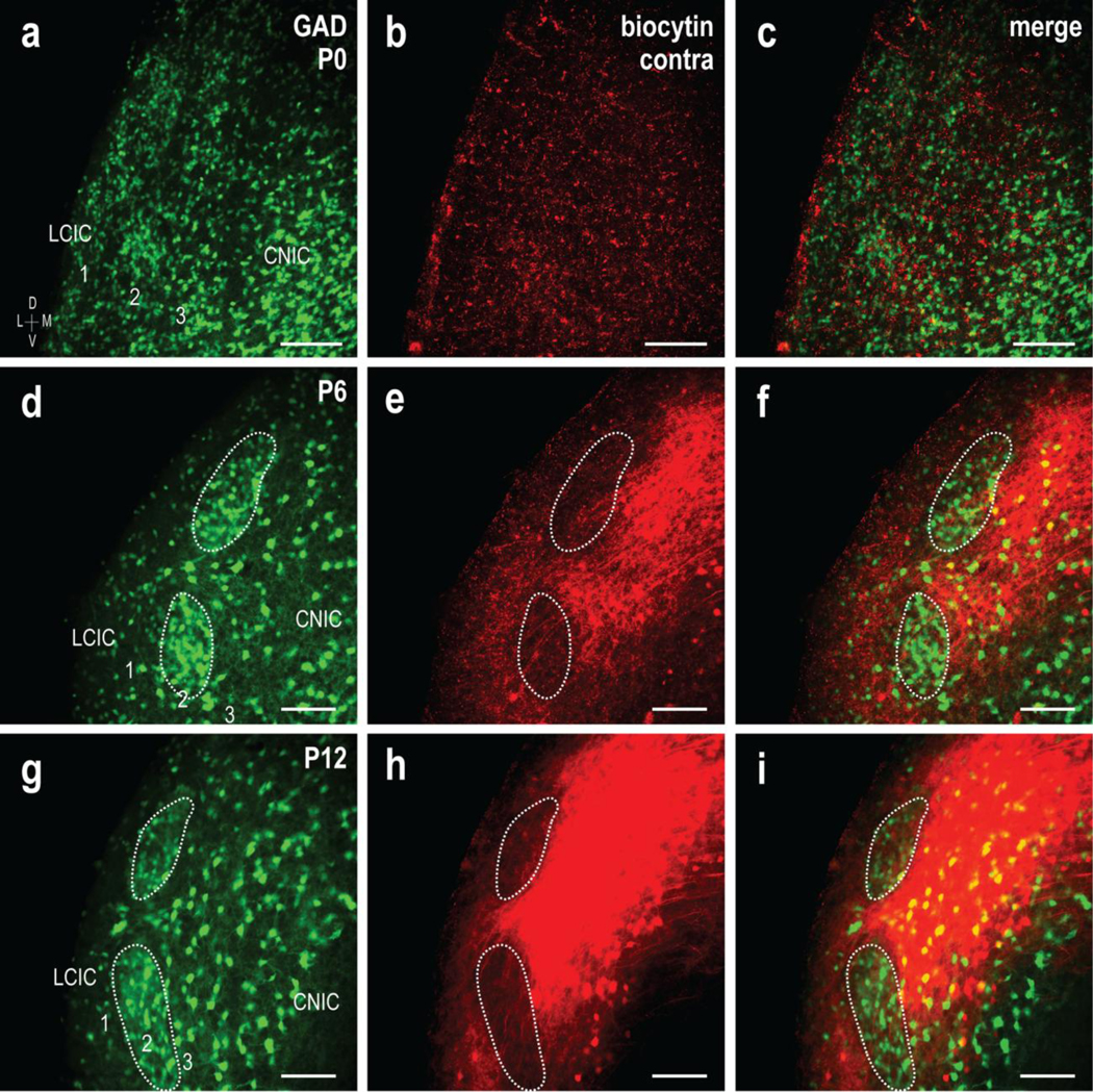
Fig 5.

Shaping of the crossed CNIC projection to LCIC extramodular zones. GAD67-GFP (green) and biocytin labeling (red) of the contralateral input at P0 (a-c), P6 (d-f), and P12 (g-i). Similar to that described for the uncrossed input, GAD modules were indistinct at birth, and while present in the contralateral LCIC, crossed projections were sparse and unorganized. Discrete GAD modules are readily identifiable at P6, with crossed inputs that appear to preferentially target surrounding extramodular zones However, compared to the uncrossed input, the crossed projection at this age across cases appeared qualitatively to have more fibers remaining within modular confines (d-f, dashed contours). At P12, the projection distribution terminated heavily within the LCIC, with dense extramodular patterns encompassing GAD-defined modules (g-i, dashed contours). Scale bars = 100μm
