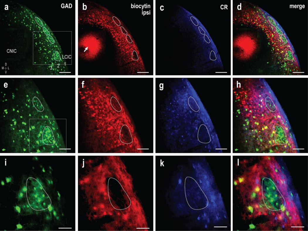
Fig 8.

CNIC targeting of ipsilateral LCIC extramodular domains in a P12 GAD67-GFP mouse. Magnification series of separate channels (GAD = green, biocytin = red, CR =blue) and corresponding digital merges. Inset boxes in (a, e) are shown at higher magnification in (e-h) and (i-l) respectively. A series of GAD modules are evident at low magnification (a, dashed contours) that appear complementary to ipsilateral terminal labeling as well as calretinin expression (a-d). Overlap of the ipsilateral input with CR-defined extramodular regions and offset from GAD modules is shown at higher magnifications (e-h and i-l). Retrogradely labeled cell bodies were frequently observed in deep aspects of the LCIC, which is keeping with recent studies of intracollicular connections in gerbil that provide evidence of bilateral LCIC projection components to the CNIC. CNIC biocytin placement in (b, arrow). Scale bars in (a-d) = 200 μm, (e-h) = 100 μm, and (i-l) = 50 μm
