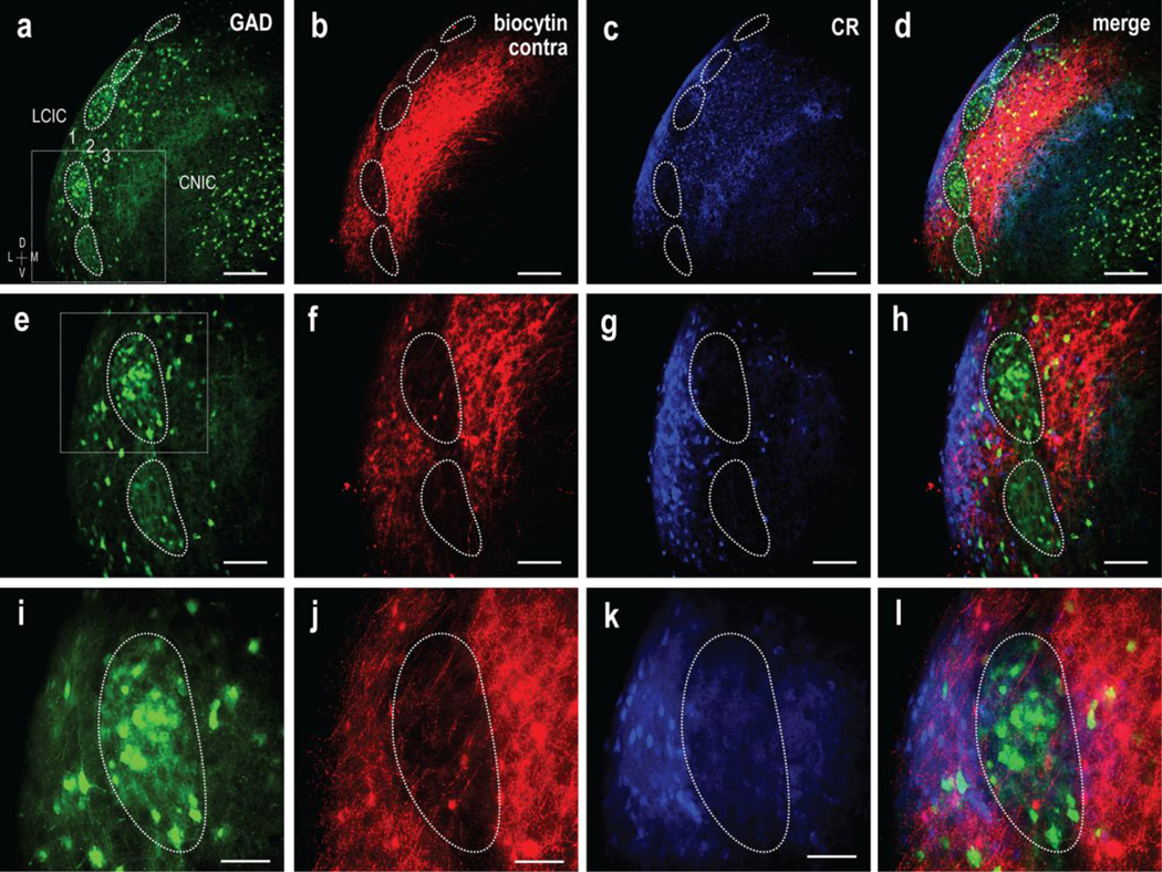
Fig 9.

Projection specificity of contralateral CNIC inputs to extramodular LCIC domains in a P12 GAD67-GFP mouse. Magnification series of separate channels (GAD = green, biocytin = red, CR =blue) and corresponding digital merges. Inset boxes in (a, e) are shown at higher magnification in (e-h) and (i-l) respectively. Similar to that described for the ipsilateral input, a series of GAD modules are evident at low magnification (a, dashed contours) that appear complementary to the contralateral terminal plexus and calretinin labeling (a-d). Alignment of the crossed projection distribution with CR-positive extramodular zones that is complementary to GAD labeling is increasingly evident at higher magnification (e-h and i-l). Scale bars in (a-d) = 200 μm, (e-h) = 100 μm, and (i-l) = 50 μm
