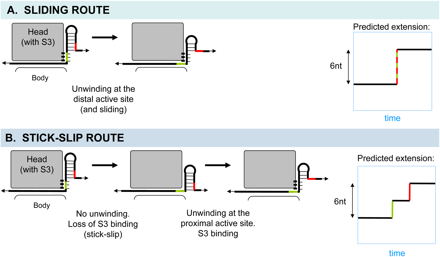
Fig. 10. Prediction of end-to-end mRNA extension in optical tweezers experiments based on the model.

A and B. The head domain of the small subunit is shown in gray, and the binding between the proximal segment of mRNA and S3 is indicated. A. The sliding route generates a major 6-nt step due to unwinding of the distal segment during translocation. B. The stick-slip route can generate two distinct ~3-nt steps, one due to slip itself during translocation, and the other due to unwinding and equilibrium binding of the new proximal segment after translocation. The ratio of the two substep sizes depends on the angle of the S3-mRNA interface with respect to the pulling force. Also see Supp. Note 5.
