
Fig. 2.
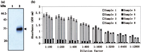
Analysis of antigenicity of expressed M protein. (a) Western blot analysis on the purified M fusion protein with the sera of SARS-CoV-positive patients. M protein was purified from lysates of BL21 cells transformed with pSARS-M plasmid through Glutathione Sepharose 4B affinity chromotography column. Purified proteins were subjected to SDS-PAGE and subsequently transferred to PVDF membrane for Western blot with the sera of SARS-CoV-positive patients. Lane 1: eluted protein of IPTG-induced BL21 transformed with the empty plasmid pGEX-6P-1 (control); Lane 2: eluted protein of IPTG-induced BL21 transformed with the recombinant pSARS-M construct. Results are Western blot probed with the serum from sample 1. Additional three experiments with the sera from sample 2, 3 and 4, respectively, were also performed and gave similar results. The properties of sera from four SARS-CoV-positive patients were listed in Table 1. Arrow indicates purified M fusion protein immunoblotting with the serum of SARS-CoV-positive patient. (b) Detection of eight human sera by indirect ELISA by using purified M fusion protein as antigen. The purified recombinant M protein was diluted and used to coat 96-well plates. SARS-CoV-positive human serum diluted in a twofold series from 1:100 to 1:12,800 was added into each well in duplicate. The OD value was read at 450 nm in CliniBio 128C reader and the cut-off value was defined as the mean OD plus 2.1 standard deviations calculated from the four negative samples used as controls. Sample 1–4 were sera from four confirmed SARS patients and 5–8 from four healthy people (controls), whose source and properties were listed in Table 1