
Fig. 3.
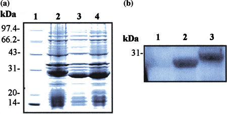
Localization of B-cell antigenic epitope in M protein. (a) SDS-PAGE analysis of truncated M protein expressed in E. coli BL21. BL21 was transformed with two truncated recombinant M protein-coding genes constructs, followed by induction with IPTG. The bacterial cultural crude materials were detected by the 10% SDS-PAGE and then stained with Coomassie blue. Lane 1: molecular weight marker; Lane 2: cellular extracts of IPTG-induced BL21 transformed with the recombinant pSARS-M construct (control); Lane 3: cellular extracts of IPTG-induced BL21 transformed with the recombinant pSARS-Ml construct; Lane 4: cellular extracts of IPTG-induced BL21 transformed with the recombinant pSARS-M2 construct. (b) Localization of B-cell antigenic epitope in M protein by Western blot with the sera of SARS-CoV-positive patients. The crude materials of inductive culture of E. coli BL21 were subjected to SDS-PAGE and subsequently transferred to PVDF membrane for Western blot analysis with the SARS-CoV-positive human serum. Lane 1: total cellular extracts of IPTG-induced BL21 transformed with pSARS-M2 construct; Lane 2: total cellular extracts of IPTG-induced BL21 transformed with pSARS M1 construct; Lane 3: total cellular extracts of IPTG-induced BL21 transformed with pSARS-M construct (control). A representative experiment is shown with the serum from sample 1. Additional three experiments with the sera from sample 2, 3 and 4, respectively, were also performed and gave similar results. The properties of sera from four-SARS-CoV-positive patients were listed in Table 1