
Fig. 4.
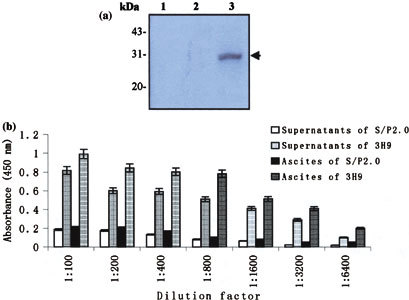
Identification of the MAb specificity. (a) Western blot analysis of the expressed M fusion protein with the MAb, 3H9. The crude materials of inductive culture of E. coli BL21 were subjected to SDS-PAGE and subsequently transferred to PVDF membrane for Western blot with the MAb, 3H9. Lane 1: total cellular extracts of IPTG-induced BL21 (control); Lane 2: total cellular extracts of IPTG-induced BL21 transformed with pGEX-6p-l (control); Lane 3: total cellular extracts of IPTG-induced BL21 transformed with the recombinant pSARS-M construct. Arrow indicates expressed M fusion protein immunoblotting with the MAb. (b) ELISA reactivity of the MAb, 3H9, to viral protein of SARS-CoV. A total of 96-well plates from the diagnostic kit detecting antibody specific to SARS virus, which were coated with the UV-irradiated and extracted viral protein of SARS-CoV, were used to determine the titers of MAb in the supernatants and ascites fluid by indirect ELISA. Supernatants and ascites fluid from 3H9 and S/P2.0 (controls) diluted in a two-fold series from 1:100 to 1:6,400 were added into each well in duplicate, respectively. The OD value was read at 450 nm in CliniBio 128C reader and the cut-off value was defined as the mean OD plus 2.1 standard deviations calculated from the four negative samples used as controls