
Fig. 5.
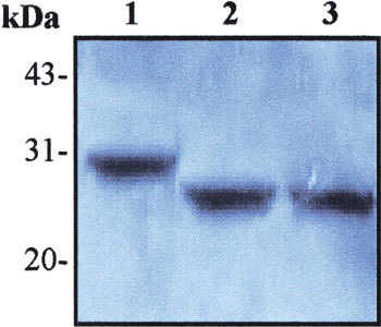
Localization of the recognizing region of the MAb, 3H9 in recombinant M protein by Western blot. The crude materials of E. coli BL21 culture were subjected to SDS-PAGE and subsequently transferred to PVDF membrane for Western blot with the MAb, 3H9. Lane 1: total cellular extracts from IPTG-induced BL21 transformed with recombinant pSARS-M construct (control); Lane 2: total cellular extracts from IPTG-induced BL21 transformed with recombinant pSARS-Ml construct; Lane 3: total cellular extracts from IPTG-induced BL21 transformed with recombinant pSARS-M2 construct