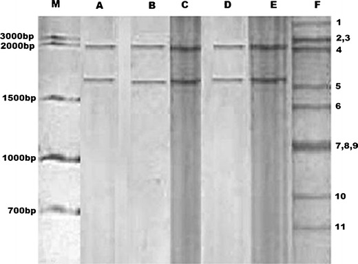
Fig. 1.

PBV electrophoretic genomic pattern obtained in faecal samples of bovine and buffalo calves. Lane indicates: M 1 kbps DNA ladder, A–E PBVs (with genomic segments of 2.4 and 1.7 kbps size), and F bovine group A rotavirus (genome segments marked 1–11 as per their gene size, segment no. 2, 3 and 7, 8, and 9 are co-migratory)
