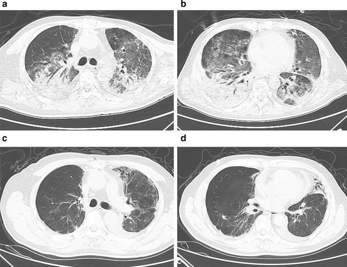
Fig. 3.

Fifty-four-year-old man with laboratory-confirmed novel influenza A (H7N9). a, b CT shows bilateral consolidation, air bronchograms, and ground-glass opacities. c, d follow-up CT scan shows obviously improvement of abnormality and occurrence of secondary fibrosis and traction, for example bronchiectasis
