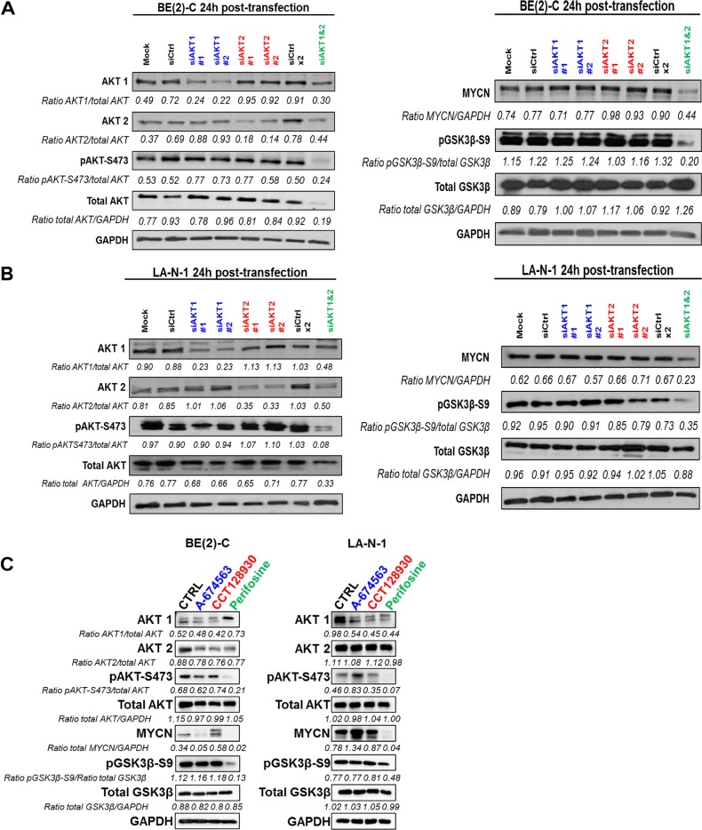
Figure 1.
Inhibition of total AKT activity suppresses MYCN expression. (A, B) Western blot analysis of AKT1 or/and AKT2 silencing in protein extracts from (A) BE(2)-C and (B) LA-N-1 cells. Cell lysates were harvested from cells 24 h after transfection with mock, siCtrl, siAKT1, or/and AKT2. Blots were probed with the indicated antibodies. GAPDH was used as a loading control. Ratios are the average of 3 independent analysis. (C) Western blot analysis of 24 h treatment of the IC50 concentration of AKT1 inhibitor (A-674563; IC50 = 0.4 and 0.25 μM in BE(2)-C and LA-N-1, respectively), AKT2 inhibitor (CCT128930; IC50 = 7 and 5 μM in BE(2)-C and LA-N-1, respectively) or pan-AKT inhibitor (perifosine; IC50 = 7.5 and 10 μM in BE(2)-C and LA-N-1, respectively) in protein extracts from BE(2)-C and LA-N-1 cells. Blots were probed with the indicated antibodies. GAPDH was used as a loading control. Ratios are the average of 2 independent analysis.

