
Figure 1.
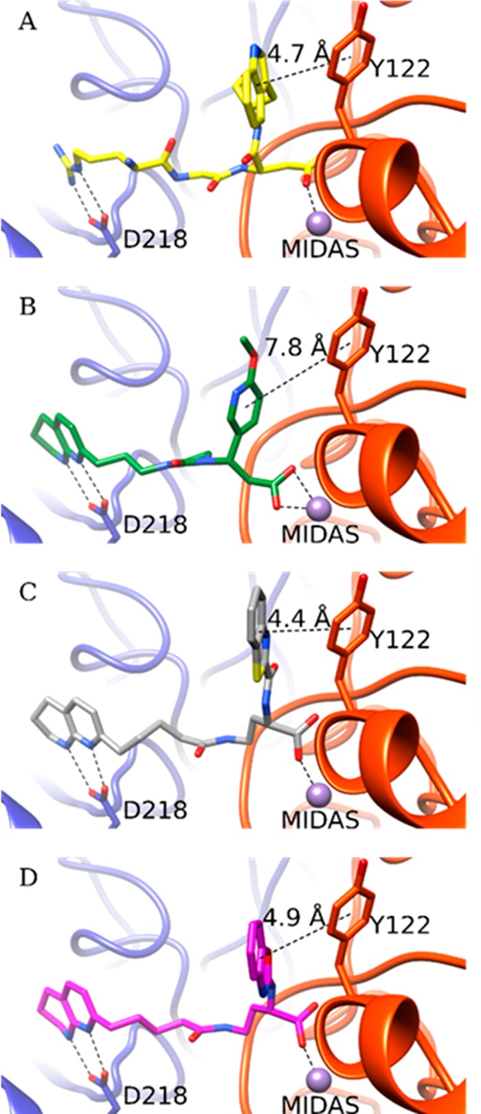
Crystal structure of the high-affinity fibronectin fragment hFN10 (A) and the predicted docking poses of MK-429 (B), TDI-4161 (C), and the S-enantiomer of TDI-3761 (D) in αVβ3. The αV and β3 backbones are shown in blue and red cartoon representations, respectively. Side chains of αV-Asp218 and β3-Tyr122 are shown as sticks. The MIDAS metal ion is shown as a purple sphere. The interactions between the compounds and αV-Asp218 and the MIDAS metal ion are indicated by dotted lines. Distances are reported in Å between the TyrR122 centroid π ring and centroids of aromatic groups at the α position of the compound’s carboxylic acid.