
Figure 5.
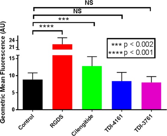
Priming of αVβ3. HEK-αVβ3 cells were either untreated (control) or incubated with 1 μM cilengitide, 100 μM RGDS, or 10 μM TDI-4161 or TDI-3761 for 20 min at room temperature, fixed with paraformaldehyde, washed, and incubated with fluorescent fibrinogen. After washing, cell-bound fluorescence was determined by flow cytometry. Compared to the control, both RGDS and cilengitide increased the amount of bound fibrinogen, whereas TDI-4161 and TDI-3761 did not. N = 7 for all values except TDI-3761, where n = 3.