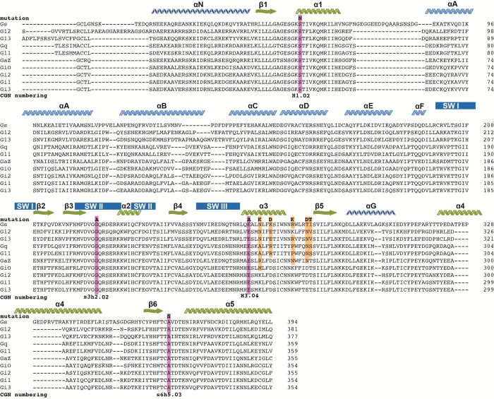
Figure 1.
Alignment of human Gα isoforms. Clustalw omega alignment of reference sequences for human Gα isoforms manually adjusted to take into account secondary elements from deposited PDB structures: 1SVK, 3FFB, 1ZCA, 1ZCB, 2BCJ, 1AZT, and 3SN6. α-Helices (zig-zags) from the α-helical domain are indicated in light blue, in dark blue are those outside either core domain and in green are those from the Ras-like domain. β-Strands are indicated in the same color scheme with wavy arrows. Secondary structure elements from the α-helical domain are indicated with letters and the Ras-like domain with numbers. The position and substitution for common DN-substitutions are highlighted in purple with the CGN numbering10 shown below. Highlighted in yellow are Gαi residues that are substituted into Gαs, which improve the dominant negative effect.

