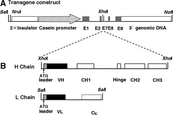
Fig. 1.

Schematic presentation of the hGHC/hGLC transgene constructs used for microinjection. a Structure of the transgene construct released from pBC1 vector with NotI/NruI for HC and NotI/SalI for LC. The transgene backbone contains 2 × chicken β-globin insulator, goat β-casein promoter, untranslated exons (E)1, parts of E2 and 7, E8, E9 and β-casein 3′ genomic DNA. The dotted lines indicate insertion of the HC and LC sequences to the unique XhoI restriction site, respectively. b Hatched boxes and solid boxes represent Ig secretory leader and the variable regions (VH and VL), respectively. Exons encoding the constant regions as open boxes are indicated in CH1, Hinge, CH2 and CH3 for HC or Cκ for LC. Translational start codons are indicated in the H chain and the L chain of Ig, respectively. Relevant restriction enzymes sites are shown
