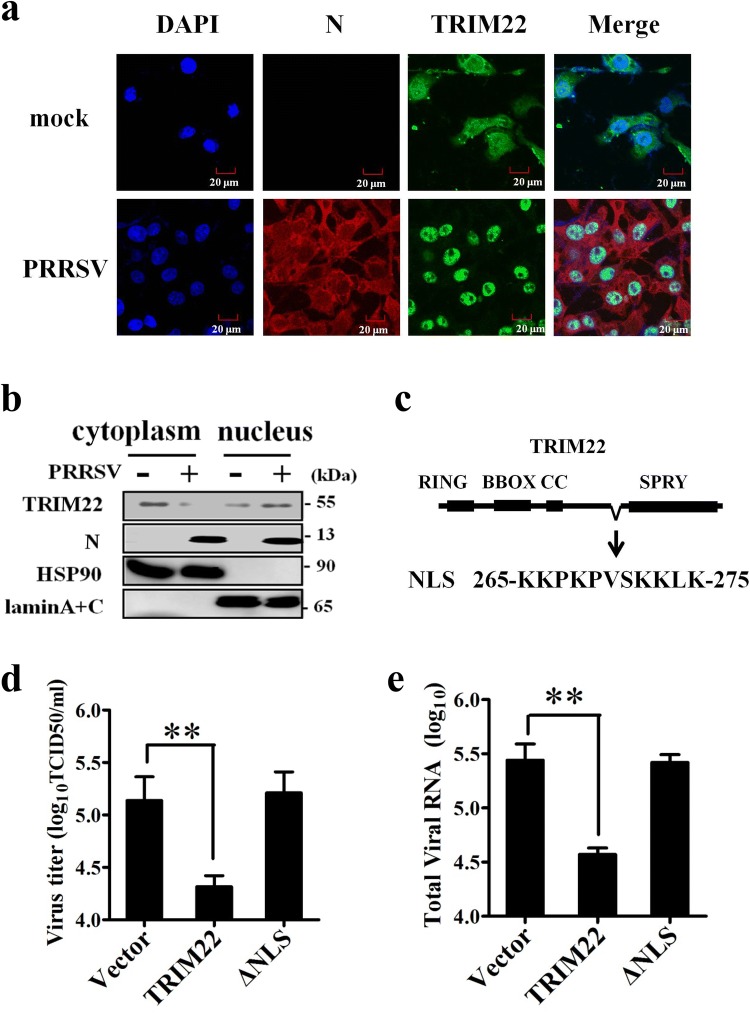
Fig. 4.
Nuclear localization signal of TRIM22 is essential for PRRSV inhibition. a Subcellular distribution of TRIM22 after PRRSV infection in MARC-145 cells. MARC-145 cells were infected with PRRSV HN1 at a MOI of 1, and were fixed for immunofluorescence analysis of TRIM22 (green), N protein (red) and nucleus marker DAPI (blue) localization. b Nuclear and cytoplasmic fractionation of MARC-145 cells infected with PRRSV for 36 h. Each nuclear and cytosolic fraction was prepared and subjected to Western blotting analysis with an antibody specific for TRIM22, LaminA + C as a nuclear protein marker, HSP90 as a cytosolic protein marker, or the PRRSV N protein. c The position and sequence of the NLS in TRIM22. MARC-145 cells transfected with Flag–TRIM22 (WT or NLS lacking mutants) were infected with 0.5 MOI of PRRSV for 48 h. d Production of progeny virus were determined using TCID50 assay. e The total cellular RNA was extracted and the mRNA levels of PRRSV N gene were determined by qPCR (Color figure online)

