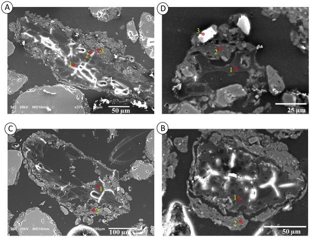
Fig 9. Scanning electron microscopy of particles of charcoal from a soil thin section collected between Layers II and III of the Bibocas II archaeological site.
The red points indicate the EDS microanalysis data shown in Table 3. Images A and B show the charcoal particles within a soil microaggregate. Image C shows a clay coating over the charcoal particles. Image D illustrates the quasi-coating formed by clay minerals and nano-charcoal particles, which is suggested by the high content of carbon in the clay matrix.

