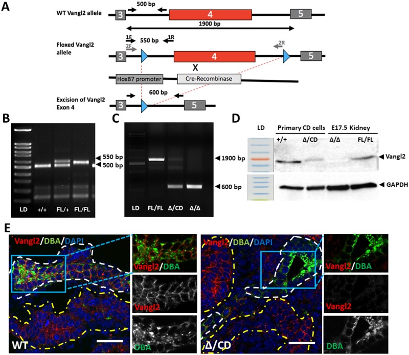
Fig 2. Generation of Vangl2 knockout mouse with excision of Exon 4.
(A) Strategy to excise Vangl2 gene in collecting duct cells using Cre-Recombinase under the HoxB7 promoter. (B) Images of tail DNA amplicons: 500 bp wildtype and 550 bp Floxed Vangl2 allele. (C) Excision of Vangl2 (Δ Exon 4) generates a 600 bp amplification fragment, the Floxed allele is amplified as a 1900 bp fragment; Fl/Fl and Δ/Δ lanes- tail DNA amplification, Δ/CD lane—amplification of DNA from medullary zone of P1 kidneys. (D) Western immunoblot with anti-Vangl2 and anti-GAPDH antibodies: protein lysates from primary collecting duct cells isolated from P5 Cre+;Vangl2+/+ and Vangl2Δ/CD mice as well as protein lysates from E17.5 kidneys of Cre-;Vangl2Fl/Fl and Vangl2Δ/ Δ were used. (E) Immunofluorescent staining with anti-Vangl2 antibody (red), DBA (collecting duct marker, green) and DAPI (blue). DBA(+) tubules are contoured in white, DBA(-) tubules—in yellow. Scale bars, 50 μm.

