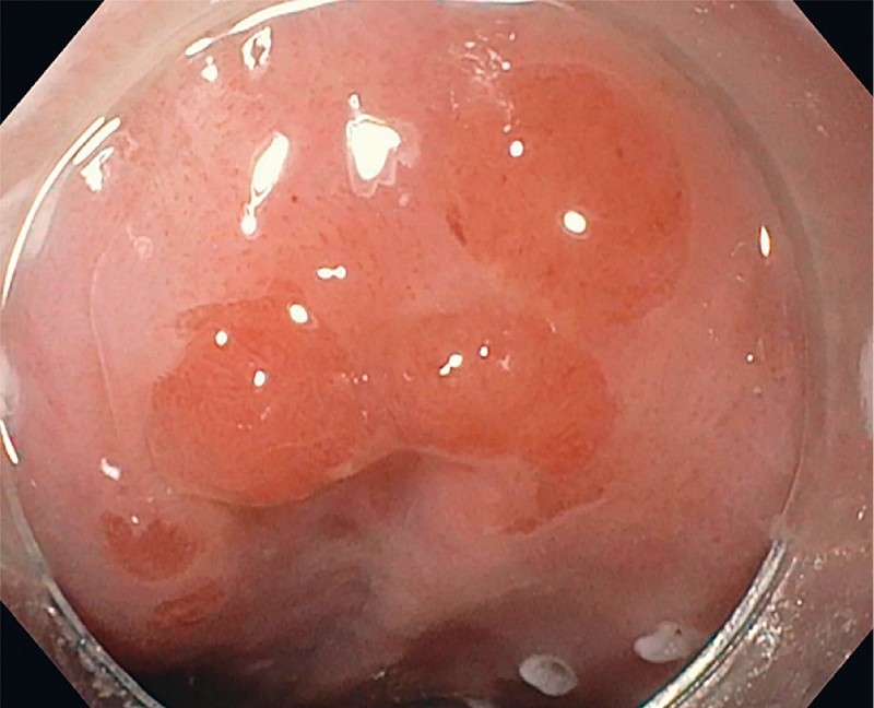
Fig. 1.

Residual nodular lesion under high-definition white light. (These images are from a 55-year-old man with BE with HGD status post-multiple RFAs. Surveillance EGD showed residual lesions with biopsy demonstrating HGD. EMR was attempted at the outside institution, however, the lesion could not be adequately raised with submucosal injection. Therefore, the patient was referred for ESD.)
