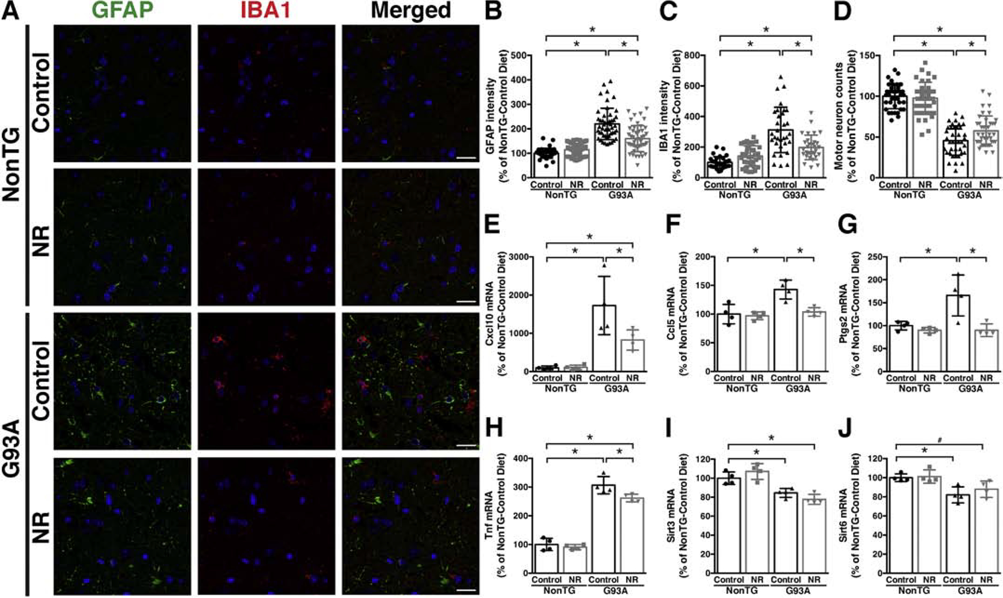
Figure 3. Dietary NR supplementation decreases glial activation and delays motor neuron loss in the spinal cord of hSOD1G93A mice.

A) Representative microphotographs showing GFAP (green) and IBA1 (red) immunofluorescence in the anterior horn of the lumbar spinal cord from early symptomatic (120 days-old) hSOD1G93A (G93A) and aged-match non-transgenic (NonTG) mice on control or nicotinamide riboside (NR)-supplemented diet. Nuclei were counterstained with DAPI. Scale bar, 20 μm. B) Quantification of relative GFAP fluorescence intensity in images from the anterior horn of the lumbar spinal cord of mice treated as in (A) (10–13 images per animal, n=4 mice per treatment group). C) Quantification of relative IBA1 fluorescence intensity in images from the anterior horn of the lumbar spinal cord of mice treated as in (A) (7–8 images per animal, 4 animals per treatment group). D) Number of large motor neurons in the ventral horn of the lumbar spinal cord of mice treated as in (A) (9–10 sections per animal, n=4 mice per treatment group). E–J) Total RNA was extracted from the lumbar spinal cord of early symptomatic (120 days-old) G93A mice and NonTG littermates on control or NR-supplemented diet and Cxcl10, Ccl5, Ptgs2, Tnf, Sirt3, and Sirt6 mRNA levels were determined by real-time PCR and corrected by Rplp0 mRNA levels (n=4 mice per group). For all panels data are expressed as percentage of NonTG-control diet mice (mean ± SD). *p<0.05 (2-way ANOVA). #p<0.05 (t-test).
