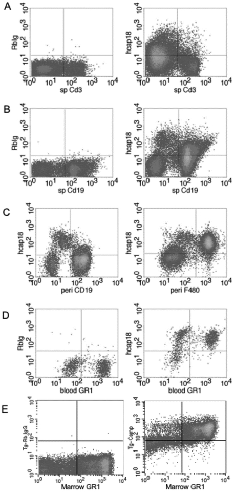
Figure 2. Tg/KO mice express hCAP18 in several lineages of immune cells.
Immune cells from three mice were pooled from spleen, peritoneal cavity, blood, and bone marrow and dual stained with lineage markers and either control rabbit IgG (left panels A, B, D, E) or anti-hCAP18 antibody (left panel C and all right panels) and analyzed by flow cytometry. (A) Splenocytes stained with the T-cell marker CD3. (B) Splenocytes stained with the B-cell marker CD19. (C) Peritoneal cells stained with CD19 (left panel) or F4/80 (right panel). (D) Blood cells stained with the granulocyte marker GR1. (E) Bone marrow cells stained with GR1. Representative plots from three different experiments are shown.

