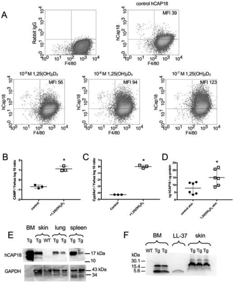
Figure 5. Induction of CAMP gene and hCAP18/LL-37 expression with 1,25(OH)2D3 treatment.
(A) BM-derived macrophages from Tg/KO mice were treated 48 h in vitro with increasing concentrations (10−9, 10−8 and 10−7 M) of 1,25(OH)2D3. hCAP18 and F4/80 staining cells (macrophages) were detected by flow cytometry. Mean fluorescent intensity (MFI) of hCAP18 is noted in the upper right quadrant. Graphs are representative of four experiments. (B-F) Dorsal skin of Tg/KO mice was treated with either vehicle control or 1 nmole 1,25(OH)2D3 for 24 h (mRNA) or 48 h (protein). Expression of CAMP (B) and Cyp24a1 (C) mRNAs (n=3 mice) determined by qRT-PCR was normalized to the housekeeping gene Ywhaz. The ratio was log10 transformed and plotted as the mean (+SD). D) hCAP18 protein levels were determined by ELISA (n=6 mice per treatment) and plotted as the mean (ng hCAP18/mg of total protein, ±SD). (E) hCAP18 expression in tissues from untreated Tg/KO and WT mice was analyzed by Western blot using 50 g protein per lane and an anti-hCAP18 polyclonal antibody. The 18-kDa pro-protein was detected in BM, skin, lung and spleen of the Tg/KO mice. The 14-kDa processed cathelin-domain was detected in the BM and skin. No hCAP18 was detected in the WT skin. GAPDH was used as a loading control, except this particular antibody does not identify GAPDH in bone marrow. (F) BM (untreated) and skin (from treated mice, panel C) samples from WT and Tg/KO mice were analyzed by Western blot with an anti-LL-37 monoclonal antibody. Synthetic LL-37 peptide (4-kDa) was included as a positive control. Both the 18- and 4-kDa forms were detected in the BM. In skin, primarily the 18-kDa was detected with very faint 4-kDa bands. Each Western blot lane represents an individual mouse. Asterisks denote statistical significance (p<0.05) determined by a two-tailed T-test.

