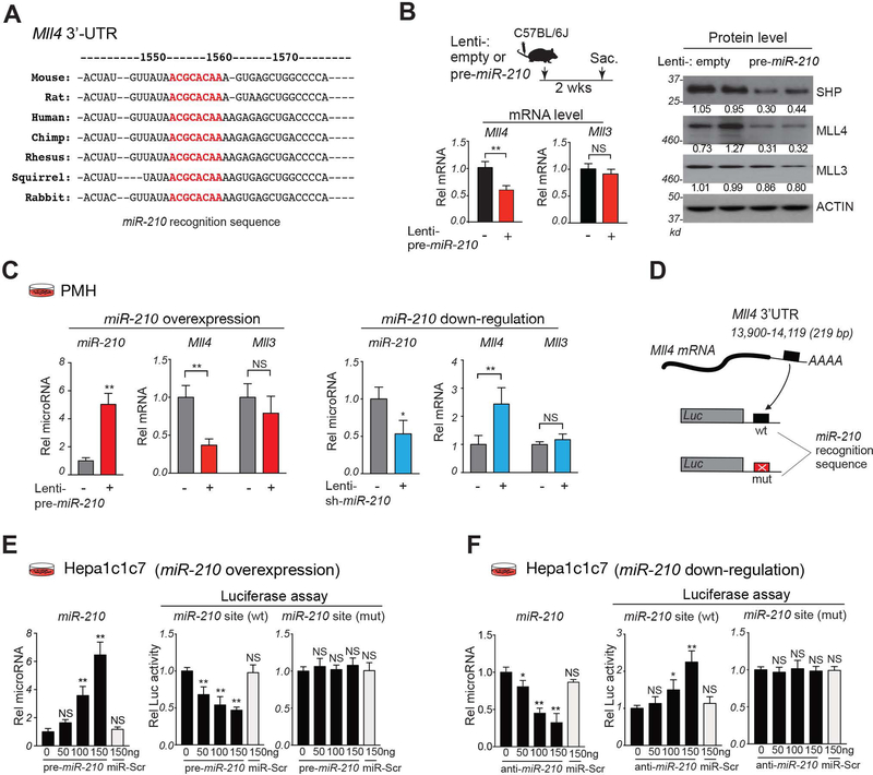
Figure 3. MiR-210 directly targets the MLL4 3’-UTR.
(A) MiR-210 recognition sequences (red) in the 3’-UTR of mammalian Mll4 mRNAs. (B) C57BL/6 mice were treated as described in the Fig. 2A legend. Experimental approach (top, left). Relative levels of Mll3 and Mll4 mRNAs with lenti-empty values set to 1 (bottom, left) and protein levels (right). (C) PMH were infected Lenti-empty (−), Lentiviruses expressing pre-miR-210 (left) or sh-miR-210 (right) for 3 days. Relative levels of miR-210 and Mll4 and Mll3 mRNA with the control RNA value set to 1. (D-F) (D) Schematic of the luciferase constructs containing WT or mutated miR-210 sites in the Mll4 3’-UTR. Hepa1c1c7 cells were co-transfected with indicated luciferase plasmids and oligonucleotides for pre-miR-210 (E), antisense-miR-210 (F), or control scrambled RNA (miR-Scr), and 48 h later, luciferase and β-galactosidase activities were measured. (E,F) Relative levels of miR-210 (left) and relative luciferase activity normalized to β-galactosidase activity (right). (B-F) Statistical significance was determined by the Student’s t-test (B-C) or one-way ANOVA (E, F), SD (n=4–5), * p < 0.05, ** p < 0.01, NS, not significant.

