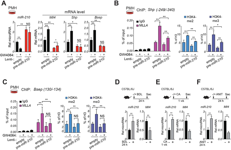
Figure 4. (A-C) MiR-210 blocks MLL4-mediated FXR-mediated induction of Shp and Bsep.
(A-C) PMH were infected either empty lentivirus or lentivirus expressing pre-miR-210 and after 3 days were treated with vehicle or GW4064 (200 nM) for 6 h. (A) Levels of miR-210 and the indicated mRNAs detected by RT-qPCR. (B,C) Occupancy of MLL4 and levels of H3K4-me2, and H3K4-me3 at the Shp (B) or Bsep (C) promoters determined by ChIP assay. (A-C) The mean and standard deviation of the mean are shown. (D-F) In cholestatic mouse models, hepatic miR-210 levels are elevated and MLL4 levels decreased. Cholestatic mice were induced by bile duct ligation (BDL) for 24 h (D), by feeding 1% CA-chow for 7 days (E), or by treatment with 75 mg/kg ANIT by gavage for 24 h (F). Hepatic miR-210 and Mll4 mRNA levels, as indicated. Statistical significance was determined by two-way ANOVA (A-C) or Student’s t-test (D-F), SD (n=4–5), * p < 0.05, ** p < 0.01, NS, not significant.

