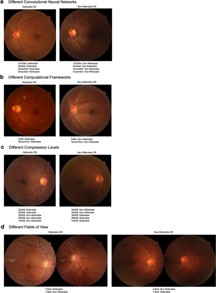
Fig. 2. Retinal image examples.
a Our results showed that using different CNNs show complementary classification of referable or non-referable DR, and these two images exhibit this agreement. b Using either computational framework similarly does not affect performance significantly as many images such as those depicted above are correctly classified as non-referable or referable DR by either framework. c Altering the image compression level does affect the DL model’s performance significantly beyond the threshold of 250 KB with a drop in sensitivity and specificity. These two photographs illustrate examples where a referable DR image is correctly identified as referable by the DL model when mild compression is introduced (i.e., a true positive case), but with further compression beyond 250 KB, this is misclassified as non-referable (i.e., a false negative case). This supports the drop in sensitivity beyond the 250 KB threshold. Similarly, this is demonstrated for a case of non-referable DR, where higher compression of the image causes a previously correctly classified image to subsequently be incorrect (i.e., a previously true negative result, now falsely classified as positive with disease), supporting the drop in specificity. d Another amendment to the image characteristics, in this case the field of view, showed reduced sensitivity and specificity when using 1-field instead of 2-field images. This example of referable DR had significant lesions present in the inferior-nasal quadrant, which were likely to be missed if using simply a macula-centered image, supporting the drop in sensitivity with the solitary use of 1-field images. Conversely, this example of healthy retina captured some dust particles in the superior and inferior nasal quadrant that might have inadvertently been misinterpreted by the DL algorithm as a lesion, prompting the misclassification as referable DR, thus supporting the drop in specificity.

