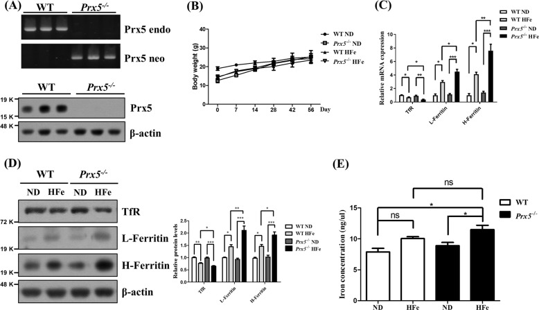
Fig. 1. Prx5 deficiency exacerbated iron overload in the hippocampus.
a Genotyping of Prx5 genes (upper panel) and the protein levels of Prx5 (bottom panel) in WT and Prx5−/− mice (n = 3). b The graph shows the body weight of WT and Prx5−/− mice (n = 6) with or without a high iron diet (HFe). c The mRNA levels of H-ferritin, L-ferritin, and transferrin receptor were evaluated by RT-qPCR in hippocampal tissues of WT and Prx5−/− mice (n = 4) with or without HFe. d The expression levels of H-ferritin, L-ferritin, and transferrin receptor were evaluated by western blot analysis in hippocampal tissues of WT and Prx5−/− mice (n = 3) with or without HFe. The graph shows the quantification of protein/β-actin. e The levels of iron concentration were measured by an iron assay kit in hippocampal tissues of WT and Prx5−/− mice (n = 4) with or without HFe. Data are expressed as mean ± SEM of three independent experiments. ns p > 0.05, *p < 0.05, **p < 0.01, and ***p < 0.001.

