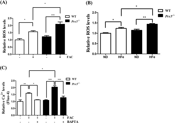
Fig. 3. Prx5 deficiency exacerbates iron overload-induced ROS production.
a Primary hippocampal neurons of WT and Prx5−/− mice (n = 4 per group) were incubated with 150 μM (49.5 μg/mL) FAC for 48 h, and intracellular ROS levels were measured by flow cytometry using CM-H2DCFDA. b Relative ROS levels were measured by OxiSelect In Vitro ROS/RNS Assay Kit in hippocampal tissues of WT and Prx5−/− mice (n = 3 per group) with or without HFe. c Primary hippocampal neurons of WT and Prx5−/− mice (n = 4 per group) were incubated with FAC for 48 h in the presence or absence of 0.25 μM BAPTA. Relative Ca2+ levels were measured by flow cytometry in primary hippocampal neurons of WT and Prx5−/− mice with Fluo-4 staining. Data are expressed as mean ± SEM of three independent experiments. *p < 0.05, **p < 0.01, and ***p < 0.001.

