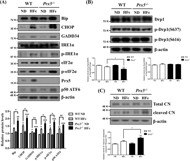
Fig. 4. Prx5 deficiency exacerbates iron overload-induced ER-stress and mitochondrial fission.
a The expression levels of Bip, CHOP, GADD34, IRE1α, phosphorylated IRE1α, eIF2α, phosphorylated eIF2α, ATF6, and Prx5 were determined by western blotting. The graph shows the quantification of protein/β-actin or phosphorylated protein/total protein. b The levels of Drp1, phosphorylated Drp1 (Ser637), and phosphorylated Drp1 (Ser616) were assessed by western blotting. The graph shows the quantification of phosphorylated Drp1/Drp1. c The level of calcineurin was determined by western blotting. The graph shows the quantification of cleaved calcineurin/total calcineurin. Data are expressed as mean ± SEM of three independent experiments. *p < 0.05, **p < 0.01, and ***p < 0.001.

