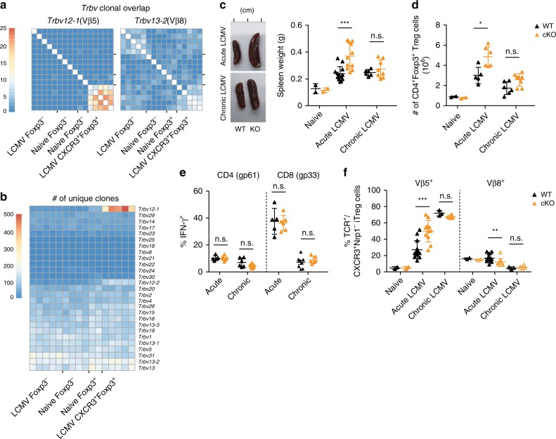
Fig. 4. CXCR3+Vβ5+ Treg cells are polyclonal.
The number of a shared clones within Trbv12-1 (left) and Trbv13-2 (right) using CD4+ T cells and b unique TCR clones within all Vβ segments was determined based on CDR3 sequences extracted from RNA sequencing data of CD4+ T cell subsets from naïve or LCMV-infected (200 f.f.u. WE) mice. Scale bars refer to the number of shared clones (a) and the number of unique clones (b). c–f Atg5fl/flxItgaxCre/− mice (cKO) and Atg5fl/fl controls (WT) were infected with LCMV WE (200 f.f.u., acute; n = 11−13), clone 13 (106 f.f.u., chronic; n = 6–8), or left naïve (n = 2). c Representative images (left) and quantification of dry weight (right) of spleens on day 14 after infection. d Total counts of splenic CD4+Foxp3+ Treg cells were determined by flow cytometry. e Splenocytes isolated on day 14 post infection were restimulated with gp61 + gp33 LCMV peptides for 4 h and IFN-γ+ frequencies were determined by flow cytometry. f Frequencies of Vβ5+ and Vβ8+ T cells among CD4+Foxp3+CXCR3+Nrp1− iTreg cells were determined by flow cytometry. Data are shown as mean ± SD; summary graphs display pooled data of 2–3 independent experiments. For statistics, Mann–Whitney U (acute) or one-way ANOVA (chronic samples) was used.

