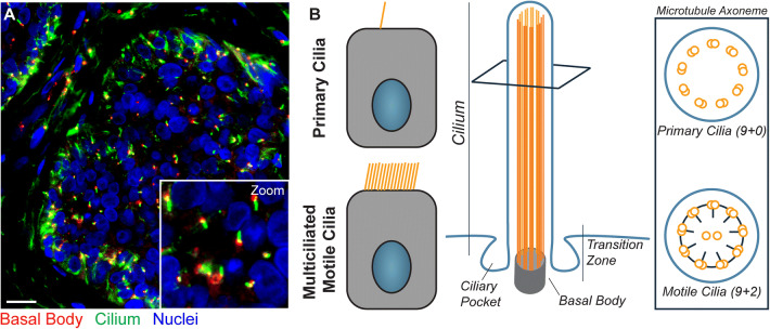
Fig. 1.
Immunofluorescence staining and structure of primary cilia. a Immunofluorescence staining of primary cilia on basal cell carcinoma is shown (scale bar 50 µm). Nuclei are highlighted in blue (DAPI), the ciliary axoneme is shown in green (acetylated alpha-tubulin), and centrioles are displayed in red (gamma-tubulin). b Unlike motile cilia, only one primary cilium is present per cell, and the primary cilium’s axoneme does not contain central microtubule singlets nor dynein arms

