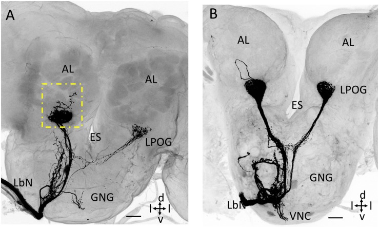
FIGURE 4.
Confocal images showing untypical staining patterns of LPO neurons in the antennal lobe (AL). (A) Maximum intensity projection showing, in addition to terminals in the LPO glomerulus (LPOG), a few branches ramifying outside the glomerulus (dotted square box). (B) Confocal image showing an axon forming a loop dorsally of LPOG. ES, esophagus; GNG, gnathal ganglion; LbN, Labial nerve; d, dorsal; v, ventral; l, lateral. Scale bars: 50 μm.

