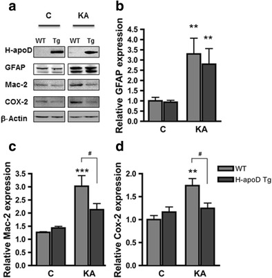
Fig. 4.

KA-induced inflammation is reduced in H-apoD Tg mice. a Western blot analysis of H-apoD, GFAP (astrocyte marker), Mac-2 (microglia marker) and COX-2 in hippocampal homogenates of vehicle (c) and KA-injected WT and H-apoD Tg mice, 3 days post-injection. β-Actin is used as loading control. Quantification of GFAP (b), Mac-2 (c) and COX-2 (d) protein expression by densitometry. Values were normalised by β-actin protein expression and by vehicle-injected WT mice values, which were given an arbitrary value of 1. Normalised values are presented as mean ± SEM (n = 3 to 5 animals for each group). Two-way ANOVA followed by Bonferroni post-test: **p < 0.01; ***p < 0.001 compared with vehicle-injected WT; # p < 0.05 compared with KA-injected WT mice. C mice injected with PBS (vehicle), KA mice injected with kainate (15 mg/kg)
