Figure.
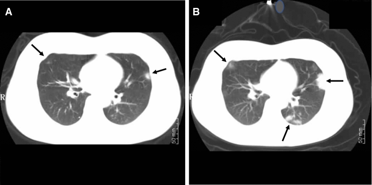
Chest computed tomography (CT) scan at the time of admission (A) of a 27-yr-old 36-week pregnant woman with coronavirus disease (COVID-19). The CT scan shows the characteristic peripheral (and/or subpleural) ground-glass opacities. These are seen in the left lower lobe/lingula junction and in the right middle lobe (arrows). Two days after admission (B), the size, density, and distribution of these opacities had progressed (arrows)

