
Figure 5.
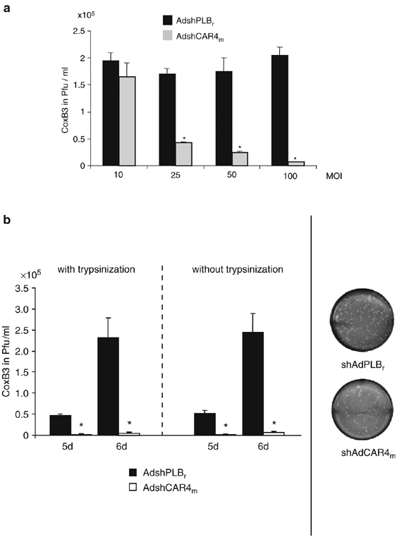
Inhibition of CoxB3 infection by mCAR silencing in HL-1 cells. (a) Inhibition of CoxB3 replication as a function of AdshCAR4m dose. HL-1 cells were transduced with AdshCAR4m or control vector at variable MOIs ranging from 10 to 100. The cells were trypsinized 24 h later, re-seeded and infected with CoxB3 (at an MOI of 1) 24 h later. Plaque assays were carried out 6 days after infection. Asterisks indicate significant differences (P<0.05). (b) Inhibition of CoxB3 replication by AdshCAR4m as a function of trypsin treatment. HL-1 cells were transduced with AdshCAR4m or control vector (MOI of 100) as in Figure 4. After 24 h, cells were trypsinized and re-seeded (with trypsinization) or the medium was changed (without trypsinization). At 48 h after transduction, the cells were infected with CoxB3 at an MOI of 1. Five and six days later, cells were lysed by three freeze/thaw cycles and virus titers were determined by plaque assay on HeLa cells (left-hand diagram). Right side: example for plaque assay on HeLa cells using a dilution of 1:500 of HL-1 cell lysate from AdshCAR4m- and control vector-transduced cells.